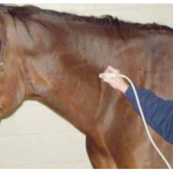
How to Perform Sonographic Examination and Ultrasound-Guided Injection of the Cervical Vertebral Facet Joints in Horses.

Introduction. Osteoarthritis of the cervical vertebral facet joints is an uncommon but documented cause of poor performance and lameness in equine athletes. Osteoarthritis of the facet joints generally occurs secondary to normal wear and tear or less frequently, to trauma. Clinical signs include neck stiffness, reluctance to bend the neck in a particular direction, and[…]

Understanding Abdominal Ultrasonography in Horses: Which Way Is Up?

Although several different types of ultrasound transducers exist, adult equine abdomens are most often imaged using a 2- to 5-MHz curvilinear array transducer. With the curvilinear transducer, ultrasound waves radiate from a curved footprint at the point of contact on the patient and generate a curved pie-shaped image through the plane of projection into the horse (FIGURE 1a, FIGURE 1b, FIGURE[…]

Evaluation of a protocol for fast localised abdominal sonography of horses (FLASH) admitted for colic.

Introduction Colic is a frequent cause of emergency calls for equine veterinarians, ranked first in importance among medical problems (TraubDargatz et al., 1991, 2001; Tinker et al., 1997). There are many causes for colic, ranging from mild to life-threatening or fatal diseases (Abutarbush et al., 2005). One of the main challenges for the equine clinician is[…]